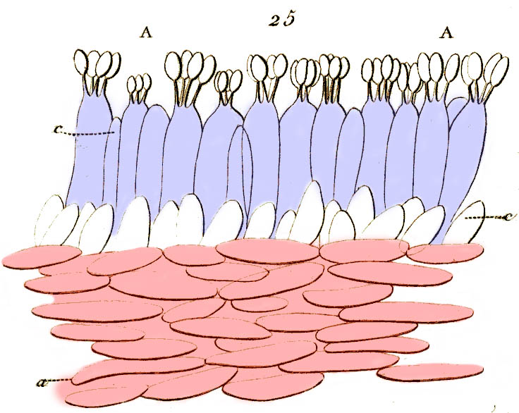
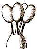
Sometimes the layer of tissue just below the hymenium has features that distinguish it both from the hymenium and from the main central trama of the hymenophore. For instance, in the cross-section of a waxy capgill in the picture, the hymenium itself is colored lilac; and the central tissue of the gill, consisting of clearly parallelhyphae, is colored pink. In between them is a layer of cells which are tilted at an angle from the central trama but not elongated and at right angles to it, like the hymenium. This layer (colored white) is the subhymenium.
The subhymenium has so far mostly been used in the taxonomy of Amanitas. Important features of the subhymenium include how many cells deep it is (only one in the diagram above), whether the subhymenial cells are bigger than the cells of the main trama, and whether the subhymenial hyphae branch before giving rise to the hymenium. Here, the subhymenium is a layer of rounded cells in between the elongated cells of the hymenium and the main trama.